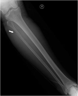

Factors that reduce bone mass had greater impact than overall health status or other risk factors for falling. Fibular fractures may also occur as the result of repetitive loading and in this case, they are referred to as stress fractures.īone mass is the key risk factor for fractures of the fibular or tibial shaft in older adults. Isolated fibular fractures contain the majority of ankle fractures in older women, occurring in approximately 1 to 2 of every 1000 white women each year. Fibular fractures in younger adults are often caused by trauma, however when compared to fibular fractures of the elderly that usually occur as a result of low energy injuries, the severity of tissue damage is equivalent. Type C: fracture above the joint level which tears the syndesmotic ligaments.Įpidemiology/Aetiology ĭistal fibular fractures represent the majority of ankle fractures.Type B: fracture at the level of the joint, with the tibiofibular ligaments usually intact.The Danis-Weber classification system uses the position of the level of the fibular fracture in its relationship to its height at the ankle joint. Type C 2 fractures result from a combination of abduction and external rotation, producing more extensive syndesmotic injury and a higher fibular fracture. There are two type C fractures: Type C 1 is an oblique medial-to-lateral fibular fracture which is caused by abduction.Type B, is caused by external rotation, it is shown as a short oblique fibular fracture directed mediolaterally upward from the tibial plafond.Type A is a transverse fibular fracture caused by adduction and internal rotation.Classification Īnkle fractures are classified according to the Danis-Weber classification system Patients may report a history of direct (motor vehicle crash or axial loading) or indirect (twisting) trauma and may complain of pain, swelling, and inability to ambulate with tibia fracture. high-energy injuries such as motor vehicle injuries, pedestrians struck by motor vehicles, and gunshot wounds.low-energy injuries such as ground level falls and athletic injuries.Mechanisms of injury for tibia-fibula fractures can be divided into 2 categories: Disruption of the syndesmosis (syndesmotic or high ankle sprain) contributes to instability of the tibiotalar joint. Stability of this ligament allows the ankle to remain stable with external rotation and during the forceful cutting movements required in many sports. The distal portion of the syndesmosis has thickened fibers to form the distal tibio-fibular ligament. The fibrous attachment between the tibia and fibula, the tibiofibular syndesmosis, prevents displacement of the lateral malleolus. Along the upper and middle lateral border of the fibula, the Peroneal (Fibularis)Longus and brevis muscles originate and provide some soft tissue protection to the fibula from direct contusion. Just below the fibular head the common peroneal nerve wraps around the fibular neck before dividing at the proximal fibula into deep and superficial branches. Proximally, the fibular head is the site of attachment of the lateral collateral ligament of the knee and of the tendon from the biceps femoris. The lateral malleolus provides key stability against excessive eversion of the ankle and foot. The fibula is a non-weight bearing bone that originates just below the lateral tibial plateau and extends distally to form the lateral malleolus, which is the portion of the fibula distal to the superior articular surface of the talus. The fibula only bears 17% of the body weight so these fractures are not as severe as weight bearing bone fractures. The fracture can happen anywhere along the fibula. Fractures of the fibula sometimes occur with severe ankle sprains. Many factors appear to contribute to the development of these fractures including changes in athletic training, specific anatomic features, decreased bone density, and diseases. 10.1.4 Phase 4- Advanced strengthening (weeks 12-16)įractures of the tibia and fibula are most common in athletes, especially runners, or non-athletes who suddenly increase their activity level.10.1.3 Phase 3- Progressive strengthening (weeks 8 to 12).10.1.2 Phase 2- Range of motion and early strengthening (weeks 6 to 8).10.1.1 Phase 1- Maximum protection (weeks 0 to 6).


 0 kommentar(er)
